The sections illustrated in Figures 31.4‒31.6 show three anatomical regions at different depths, which are differently perceived by palpation: The cerebral cortex (pallium), region of the commissures, and central nuclei. These three different levels represent parts of the brain that are different in terms of evolutionary development. These are also reflected in ontogenesis. Hence we are able to ‘grasp’ not only developmental patterns that take place over time, but also the functions associated with these regions.
Figure 31.4- The three different depths of palpation: coronal section.
Figure 31.5 - The three different depths of palpation: frontal section.
Figure 31.6 - The three different depths of palpation: sagittal section.
Palpation detects the following three growth movements:
1 Dorsal growth: It feels as if the brain is rising or descending in your palpating hands. This is the growth movement of the deepest level.
2 Radial growth: The brain expands in your palpating hands and there is a sense of distension. This is the middle level.
3 Appositional growth: It feels as if the brain is folding in your palpating hands, and the gyri are taking shape (like the growth of a cauliflower). This is the outermost level, the newest in terms of the developmental history.
Figure 31.10 - The three different growth dynamics.
1 Dorsal growth ‘The brain ascends/descends in the palpating hands’
2 Radial growth ‘The brain expands in the palpating hands’
3 Appositional growth ‘The brain bulges in the palpating hands’
Palpation of the different levels of the brain, described As we practice and develop the capacity to palpate the different levels of the brain, this enables us to comprehend the first developmental patterns of the brain. As it develops, the brain takes shape by performing various developmental movements. The levels represent different partial regions of the brain, each with different degrees of myelination, neuron density and structure. The patient lies supine and relaxed, leaving sufficient space at the head end of the treatment table for the practitioner’s forearms or elbows (depending on the particular hold being used for contact). This enables the creation of the appropriate fulcrum without any further effort during the palpation itself. If wished, you can help the patient to achieve better relaxation by placing a support under the patient’s knees, or by raising the patient’s feet.
Practitioner: Before taking up contact, center yourself as previously described (see ‘Preparation’ above).
Hand position: Place the fingers of each hand gently on each side of the parietal bones with a gentle contact. Your thumbs should be touching, above the sagittal suture, so as to create a fulcrum. (See Figures 31.7-31.9)
Once you have taken up the contact, wait to discover what information on the individual developmental patterns reaches your hands. You use this as your guide to help you perceive the different levels and their developmental dynamics. This palpation exercise prepares you to assess the influences and pivot points of brain development, to assess different regions of the brain and the patterns of relationship to other organs and developments of organs. The growth dynamics can be sensed not only at the brain, but also at the spinal cord (Figure 31.11) It is also helpful to use this form of palpation following medullary contusion. The patterns of tension shown in Figure 31.12 should be assessed in this case.
Figure 31.11 - Growth dynamics in the spinal cord.
Figure 31.12 - Patterns of tension appearing following contusion.
You need to assess when and which region the compression occurred. The following causes may account for the compression:
– Trauma affecting the adult (whiplash injury, emotional trauma)
The approach from here on is to balance the corresponding space-time patterns of tension and enable tissue release.
Palpation: position of the central nervous system (brain and spinal cord). The task here, in contrast to the assessment of growth movements, is to assess the position of the central nervous system. The following positions can be distinguished:
1 “Ascended” – “descended” brain
2 Global retroposition
3 Global anteposition
Course of palpation: The patient lies supine and relaxed, leaving sufficient space at the head end of the treatment table for the practitioner’s forearms or elbows (depending on the particular hold being used for contact). This enables the creation of the appropriate fulcrum without any further effort during the palpation itself. If wished, you can help the patient to achieve better relaxation by placing a support under the patient’s knees, or by raising the patient’s feet.
Practitioner: Before taking up contact, center yourself as previously described (see ‘Preparation’ above).
Hand position: Place the fingers of each hand gently on each side of the parietal bones with a gentle contact. Your thumbs should be touching, above the sagittal suture, so as to create a fulcrum. (See Figures 31.7-31.9)
Once you have taken up the particular contact, wait to discover what information on the position of the central nervous system reaches your hands. The following positions – illustrated below – can be found. (The Figures represent an idealized form. Various kinds of rotation and lateral variation are also possible.) In embryological development, the CNS executes developmental movements as demonstrated (for example) by Blechschmidt. Palpation should assess what information we receive about the brain and spinal cord regarding their position. All these positions are associated with certain hinge points, around which developments of movement take place. As palpation proceeds, we can build up fulcrums relating to these points and establish relationships with other developments of organs within the body as well as with emotional states.
Van den Heede P, Goldenstein R, Liem T. The brain as a morphogenetic field. In: Liem T. Van den Heede P. Foundations of Morphodynamics in Osteopathy: An Integrative Approach to Cranium, Nervous System, and Emotions, 2017; 1. Aufl. Handspring Publishing Limited, Pencaitland.
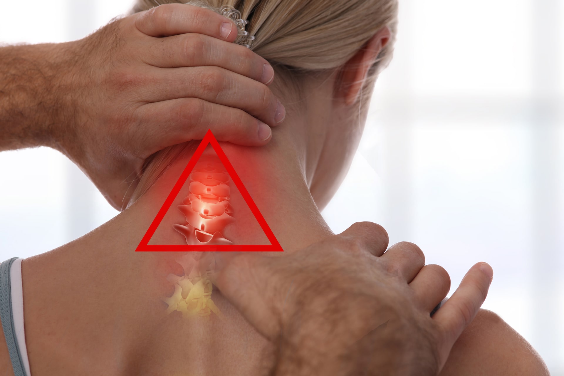
Osteopathische Interventionen können – laut einer Studie von 2022 – bei Erwachsenen mit unspezifischen Nackenschmerzen zu einer Verbesserung des Schmerzniveaus und des funktionellen Status führen.
Entdecken Sie 11 praktische Tipps, um eine dauerhafte Immunaktivierung zu vermeiden und Ihre Gesundheit zu fördern, von regelmäßigen Bewegungspausen bis hin zur richtigen Ernährung.
Osteopathische Interventionen können – laut einer Studie von 2022 - bei Erwachsenen mit unspezifischen Nackenschmerzen zu einer Verbesserung des Schmerzniveaus und des funktionellen Status führen. ...