On various occasions at the end of a course on the cranial concept, Sutherland liked to hold a lecture entitled “A tour of the minnow.”8 The underlying idea was to illustrate insights into and about the living brain. I adopted Sutherland’s idea, and we will accompany the minnow on a sightseeing tour to the period when the eye is being formed. This description will serve as an example of fluid developmental dynamics. By palpation using gentle and meaningful hand contact, you can attempt to follow these developmental dynamics:
- The minnow finds itself in the primary forebrain.
- It observes how the primordium of the eye appears on day 25/26 (at a time when the neural tube has not yet closed) as a small groove (the optic sulcus) on the forebrain.
- It accompanies this development further and notes how this groove rapidly deepens and how, following closure of the neural tube, the tiny optic vesicle appears as a projection of the primary forebrain.
- The minnow attentively observes how the optic vesicle in its further growth dynamics reaches toward the external boundary of the body on about day 32 and finally touches it.
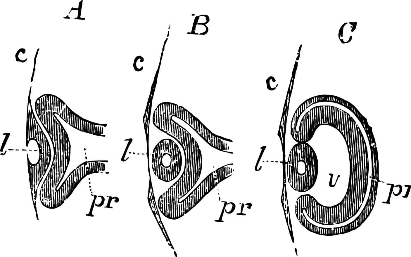
- As soon as it touches the epidermis, the optic vesicle flattens and undergoes indentation to form the optic cup. The minnow witnesses a repeat of developmental dynamic movements similar to those leading to the formation of the neural tube.
- At this point of contact with the outer body surface something else occurs. The minnow again notices how the initially flat epidermal cells become columnar, and undergo condensation and invagination. This developmental dynamic is similar to gastrulation and neurulation. The optic vesicle is pressed in by the future lens material and yields around it.
- The minnow accompanies the lens on its inward migration until this finally detaches from the skin surface. (The process is similar to the formation of the neural tube from the neural plate, except that in this instance a circular rather than a cylindrical basic form develops.)
- On about day 40 the lens is surrounded by the optic cup that has formed.
- On the inner surface of the retina the cell layer has developed to form neurons, the axons of which grow across the inner surface of the retina and finally into the optic stalk. The minnow allows itself to be carried to an outlet for the stream carrying it, to the exit point for the optic nerve, and reaches the midbrain where the axons spread into the midbrain roof.
- Finally, the minnow emerges in the central fovea, as if carried along by the current of a stream.